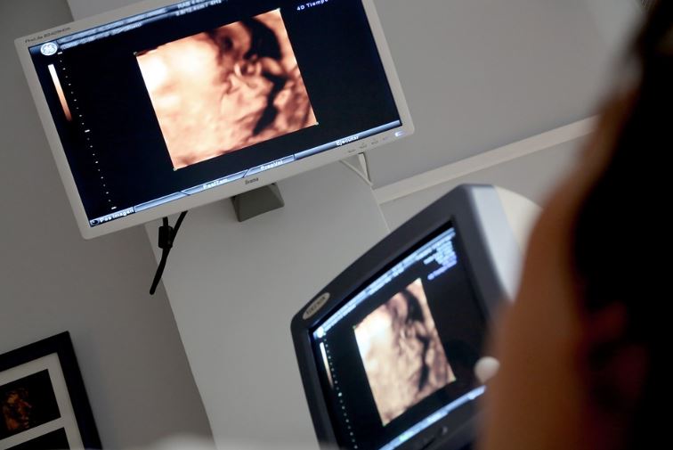
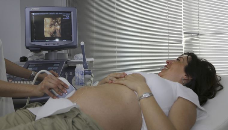
An ultrasound machine is a device that shows us pictures or video footage of the inner parts of the human body. During an ultrasoud scan, many pregnant mothers see their offspring for the first time. These scanners work by using high-frequency sound waves to create images of the interior of the uterus. The ultrasound machine’s pictures are captured by computer software.
There are numerous reasons to use ultrasound technology over other types of imaging technology, which include the provision of real-time visuals of organs or the fetus. Ultrasound scans are non invasive and do not use ionizing radiation substances which can harm the embryo or fetus. During the early stage of a woman’s pregnancy, she is carrying an embryo. When all its limbs and organs are formed, i.e., by the 11th week of pregnancy, it is a fetus.
The two best forms of ultrasound technology are 3D and 4D scans. What is the difference between a 3D and $D scan? Let’s see:
What is a 3D Ultrasound Machine?
3D ultrasound technology is an evolution of 2D that mainly provides a better image. When using one of these machines, you will be provided with a 3D image that’s composited from sound waves that are taken at several different angles.
The volume data that allows for the capture of 3D images is acquired and analyzed before being displayed. If you’re using this type of scanner, the array sensors within the machine only require a single pass to obtain the necessary data. During this pass, a large number of 2D frames are captured rapidly.
A computer gathers all the data from these 2D scans and creates a three-dimensional or 3D image that humans can see and understand.
Benefits of a 3D Ultrasound
There are many advantages to using a 3D ultrasound machine over a 2D one. For instance, the usage of virtual planes allows for enhanced visualization of heart structures in the fetus, which makes it easier to accurately detect birth defects.
With a 3D scan, health care professionals can more easily and accurately detect skeletal and facial defects. There’s also a reduced need for operator skill for the diagnosis of fetal anomalies.
What is a 4D Ultrasound Machine?
The four dimensions are: 1. Width. 2. Height. 3. Depth. 4. Time. 4D scans differ from their 3D counterparts in the same way videos differ from still photographs. With a 4D scan, the health care professional can see a real-time streaming video of the embryo, fetus, or target organ. In other words, it is an ultrasound video.
While 3D ultrasounds improve over 2D ultrasounds by allowing for the capture of static 3D images, 4D portable ultrasounds are able to display movement. As such, the health care professional who administers the scan can view, for example, blood flow to the heart valves of the fetus.
Parents can see their offspring kicking, stretching, yawning, and even opening and closing their eyes. Seeing them for the first time is an amazing moment. Seeing the fetus moving is mind-blowingly bonding event.
Benefits of a 4D Ultrasound
The only difference between a 3D ultrasound and a 4D ultrasound is that the latter provides a video of the static image that’s delivered by a 3D ultrasound. As such, most of the benefits of a 4D ultrasound are the same as a 3D version.
When using this technology, less time is required for a fetal heart screening or to be able to spot a defect.
Many pregnant women who undergo a 4D ultrasound scan will also exhibit healthier behaviors because they were able to view their baby in real-time. By capturing a video of the ultrasound image, this type of technology allows for more precise identification of a wide range of fetal anomalies, such as defects around the heart, face, neural tube, limbs, and skeleton.
4D scans also have all the benefits of 2D ultrasounds, which include everything from being able to view how much the fetus is growing to hearing and observing its heartbeat.
Final Thoughts
For the pregnant mother, her first ultrasound scan is typically one of the most exciting and emotional events in her life. She meets her baby for the first time.
Remember that a mother will only be able to see her baby’s face if they are facing her belly button. If the fetus’ head is already positioned in her pelvis, it might not be possible to get a view of the face.